Mitosis Model, Dimensions: approx. 60x40x6 cm3, Advantages: Chromosomes coloured according to modified AZAN staining colours, Cell components are colour-coded in accordance with educational aspects, Attaching magnets on the rear, Storage system, Enlarged 10, 000 times
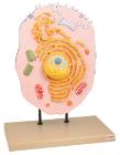
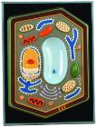
Enlarged 20,000 times. The model shows delicate structure of an animal cell. Features organelles nucleus, endoplasmic reticulum, mitochondria, ribosomes respectively polysomic and Golgi apparatus. Also showing centrioles, lysosomes and fat vacuoles.
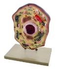
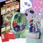
Three dimensional model showing electron microscopic structure. Organs like nucleus, Nucleolus, endoplasmic reticulum, mitochondria, ribosomes respectively polysomes and golgi apparatus. Showing centrioles, lysosomes and vacuoles.
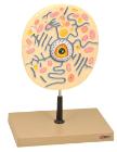
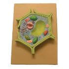
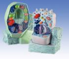
This plant cell model is separated in 4 parts. Showing details of cell wall and inner details of cell wall, nucleus is separated and defined in a 3-D way cut section of chloroplast is shown.
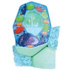
Science BioSigns Animal Cell Model, Hands-on interactive 14-piece model teaches about cell membranes and mitochondria, Provides a magnified and cross-sectioned detailing of an Animal Cell, Includes Guide
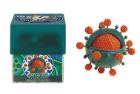
Virus Model, Hands-on interactive model provides a magnified and cross-sectioned detailing of Bacteria, Able to be separated, with both halves filled with a cross section of genetic material and flagellum, With Guide